Research
We study the structure and electrical properties of axonal excitable sites, where electrical signals are generated. These include the axon initial segment (AIS), where signals are initiated, and the nodes of Ranvier, where signals are regenerated along myelinated axons. These functions are facilitated by their unique molecular composition, enriched in ion channels.
Generation of Electrical Signals in Axons
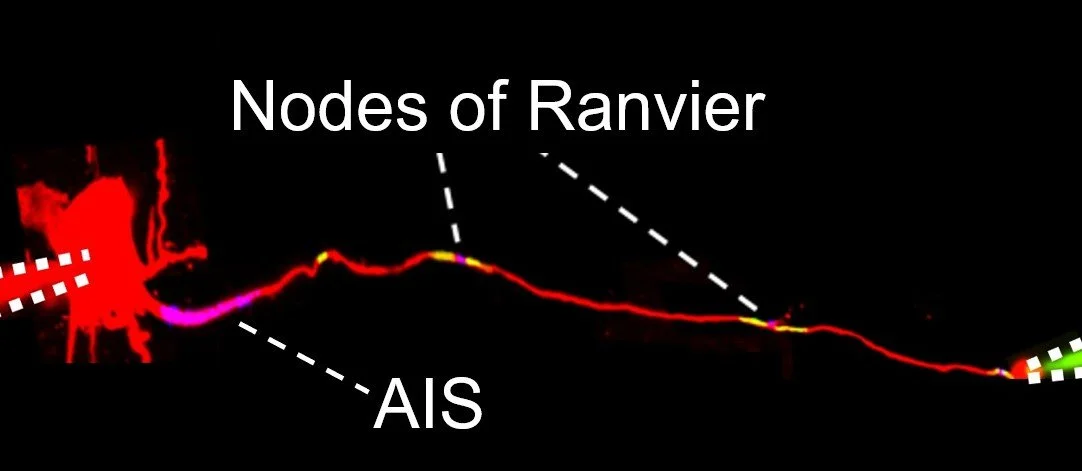
A dye-filled neuron (in red) is patch-clamped at its soma and axon end (green pipette on the right). Simultaneous electrical recordings at both sites allow us to study how electrical signals propagate along axons. The adenosine receptor A2bR (blue, appearing purple when overlapping with the axon) is expressed at both the AIS and the nodes of Ranvier (Lezmy et al. Science, 2021), and Caspr (green, appearing yellow when overlapping with the axon) labels the paranodes flanking the nodes.
Using multiple patch-clamp recordings combined with confocal and two-photon imaging (both in slices and in vivo), we investigate how electrical signals propagate along axons. We have found that adenosine, via activation of adenosine receptors expressed at the nodes and paranodes, is a key regulator of axonal conduction. We also develop computational models and experimental tools to study signal propagation in white matter.
Propagation of Nerve Signals in the White Matter
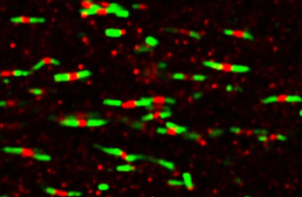
Nodes of Ranvier are found in abundance in the white matter. Here, in the corpus callosum of a mouse, they are labelled in red for the Nav sodium channels and are flanked by the Caspr-labelled paranodes in green. Node length varies in response to neuronal and astrocytic activity (Lezmy et al. BioRxiv, 2025).
Since the axonal environment is rich in glial cells, we are interested in their impact on axonal activity. We have previously found that astrocyte calcium activity near nodes of Ranvier modulates nodal structure and function. We study the roles of astrocytes, microglia, and oligodendrocyte precursor cells near axonal excitable sites and in white matter.
Regulation of Axonal Activity by Glial Cells
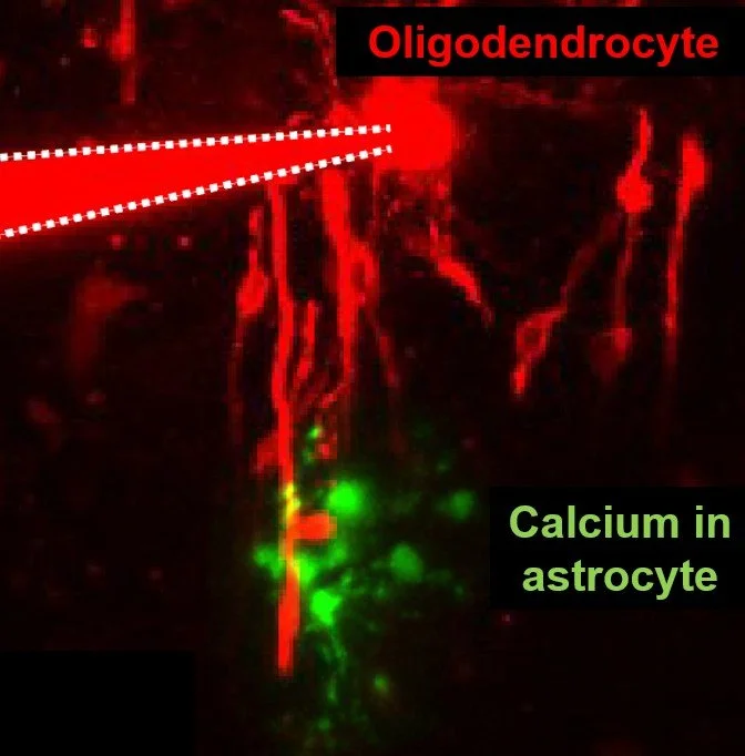
Different cell types make up the axonal environment. Here, an oligodendrocyte (patch-clamped and dye-filled in red) exposes the myelinated internodes emerging from it, which wrap axons. A nearby astrocyte is also patch-clamped to manipulate and monitor calcium activity (in green). Astrocytes regulate oligodendrocyte conductance and internode length (Lezmy et al. BioRxiv, 2025).
We investigate how the AIS and nodes of Ranvier are disrupted in multiple sclerosis and Alzheimer’s disease, and the impact of these changes on axonal conduction. We study how changes in glial activity in these diseases affect axonal excitable sites, and the impact glia have on spike propagation in the diseased brain.
Axonal Conduction in Disease Models
We thank the European Research Council (ERC), the Multiple Sclerosis Society UK and the European Molecular Biology Organization (EMBO) for their recent support of our research.